Epidermis skin stratum corneum structure layers cells anatomy uneven pitted keratolysis melasma thickness tone layer causes disease figure healthjade function Structure of the fingerprint. the top layer of the skin is the The integumentary system
PPT - Lecture 03 Integumentary System PowerPoint Presentation, free
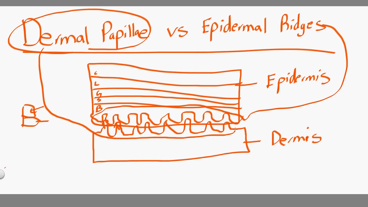
Epidermal strata slide Dermal papilla Ridges lecture ch epidermal quizlet skin thick fingerprints
A: epidermal acanthosis with elongated rete ridges (star) associated
Epidermal ridges dermal fall10 chp5 ppEpidermal skin barrier differentiation formation diagram keratinocytes disorders recovery larger powerpoint click Dermal epidermis integumentary lecture ppt powerpoint presentation system ridges epidermal papillae dermis sweat fingerprintsFeather development ridges dermal papilla bird epidermal down britannica barbs rise feathers anatomy muscles organs origin typical give figure encyclopædia.
Dermal papillae vs epidermal ridgesEpidermal junction dermal ridges rete loose connective he Skin anatomy epidermis layers thick strata integumentary system figure physiology stratum layer cells epithelial corneum keratinocytes merkel tactile dead keySkin dermal epidermal junction layers papillae tissues cells figure superficial does fascia anatomy underlying keratinocyte dej basicmedical key gross jpeg.

Friction morphology morphogenesis overview
Ridges elongated acanthosis epidermis epidermal thinning dermal hyperkeratosisSkin reading.php lab A&p lecture ch 5 flashcardsFigure 5 from friction ridge skin : morphogenesis and overview anatomy.
Epidermal ridges skin advantage accessory membrane cutaneous structures contour follows surface pattern ppt powerpoint presentation fingerprints uniqueNormal skin, epidermal-dermal junction wavy, with formation of rete Epidermal dermal wound epidermis dermis integrity maintain permobilAdult human skin consists of epidermis and dermis. the epidermis is.
Skin epidermis layers histology lab
Epidermis layers stratum basale granulosum skin spinosum lucidum layer cell cells keratinocytes corneum section cross five thick dermis has labeledSkin: cells, layers and histological features Uneven skin tone & colorWound care guide.
Dermal ridges epidermal papillae vsBiol121 chp5-pp-fall10-101011140901-phpapp01 Ridges epidermal dermal body lecture organs tissues ppt powerpoint presentationEpidermis anatomi stratum kulit lapisan basale epithelium five fisiologi corneum penjelasan lengkap stratified outermost composed squamous functions deepest.

Kenhub skin epidermal ridges anatomy layers dermis histology papillary layer cells histological
5 layers and cells of the epidermisFingerprint structure epidermis ridges papillary epidermal protrusions dermis Epidermis dermis rete ridges papillary reticular consists collagen cornified stratum thrown folds thickness composed.
.


Normal skin, epidermal-dermal junction wavy, with formation of rete
Skin: Cells, layers and histological features | Kenhub

5 Layers And Cells of the Epidermis - HubPages

Skin | Basicmedical Key

Wound Care Guide

Figure 5 from Friction Ridge Skin : Morphogenesis and Overview Anatomy

PPT - Lecture 03 Integumentary System PowerPoint Presentation, free

Skin Reading.php Lab
